

By Dr. Kevin Lam, DPM, FACFAS – Clinical Director, Family Foot & Leg Center
For millions of people suffering from chronic foot pain, a Morton’s neuroma can feel like an unending battle. Characterized by a sharp, burning sensation or the feeling of standing on a folded sock, this nerve condition often drives patients through a relentless gauntlet of treatments. From orthotic inserts and anti-inflammatory medications to corticosteroid injections, physical therapy, and even dry needling, many find themselves running out of non-invasive options. When conservative therapies fail, foot surgery is traditionally presented as the final destination.
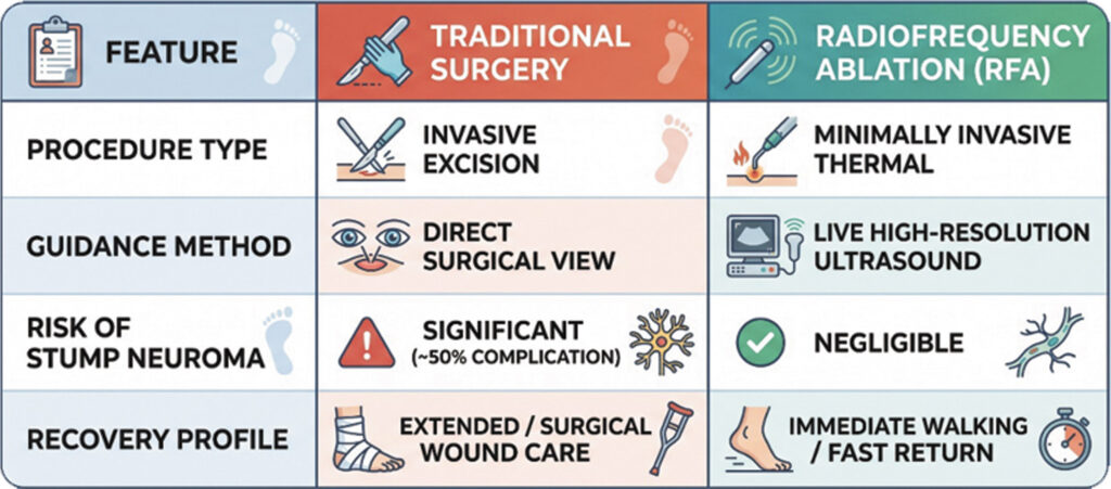
However, standard surgical neurectomy carries a critical, often unspoken risk: a 50% complication rate. Even in the hands of the most skilled surgeons, no one can accurately predict whether a patient will develop a painful stump neuroma—a complication where the severed nerve forms a new, hypersensitive bundle of scar tissue. Faced with a coin-flip chance of postsurgical complications, many patients—and informed practitioners—look for a safer, highly effective alternative.
That alternative is live, ultrasound-guided radiofrequency ablation (RFA).
Why Diagnostics Matter: MRI vs. Live Ultrasound
A common misconception in podiatric care is that a magnetic resonance imaging (MRI) scan is the gold standard for diagnosing a neuroma. In reality, MRIs provide static photos that are notoriously inaccurate for this specific condition, even when contrast dye is used. A static image cannot capture how the nerve behaves dynamically under pressure.
High-resolution ultrasound offers a dynamic, real-time diagnostic solution. By scanning the bottom of the foot between the metatarsal heads—the “knuckles” of the forefoot—a practitioner can clearly visualize the compressed nerve as a distinct nodule. Ultrasound allows the clinician to squeeze the patient’s toes together during the examination to watch the nerve move in real time. This level of direct visualization is vital for precision targeting during treatment.
How Radiofrequency Ablation Works
Radiofrequency ablation is a minimally invasive, in-office procedure designed to disrupt the pain signal sent by the inflamed nerve without physically removing it or damaging surrounding structural tissue.
The Procedure Step-by-Step:
1. Targeted Anesthesia: Rather than injecting local anesthetic blindly into the forefoot, ultrasound guidance is used to locate the nerve near the ankle first. Numbing the nerve at this site completely deadens sensation across the bottom of the foot, ensuring the patient remains entirely comfortable throughout the procedure.
2. Precision Front-Angle Access: Modern techniques avoid entering through the top of the foot. Instead, an RFA probe is inserted directly through the front space between the toes at a calculated angle. Guided by live ultrasound imaging, the clinician maneuvers the probe until it sits precisely within the affected nerve tissue.
3. Thermal Ablation: Once the probe is correctly positioned, radiofrequency energy delivers targeted heat at 90°C. This controlled thermal energy creates a specific micro-injury to the nerve, effectively turning off its ability to transmit chronic pain signals to the brain.
Clinical Outcomes and Patient Recovery
Because radiofrequency ablation does not involve surgical incisions, structural disruption, or nerve excision, it bypasses the severe risks associated with conventional surgery—most notably the formation of stump neuromas.
The typical protocol for radiofrequency ablation includes up to two targeted sessions. Clinical results from this two-step approach show substantial promise:
• Complete Resolution After One Session: A significant number of patients experience total relief after their initial procedure and do not require the second scheduled session.
• Progressive Relief: Patients who retain residual symptoms after the first treatment frequently report a 50% to 80% reduction in pain, making the second protocol treatment highly effective at resolving remaining discomfort.
So compelling is the safety and efficacy profile of RFA that podiatric specialists routinely perform the procedure on their own family members—preferring a 90°C thermal treatment over the 50% complication risk of an operating room.
A Modern Alternative to the Knife
When conservative methods fail to deliver relief from neuroma pain, patients are often told that surgery is their only remaining option. However, live ultrasound-guided radiofrequency ablation provides a proven, non-surgical path forward. By combining real-time imaging with precise thermal energy, RFA eliminates pain signals at the source, offering a high success rate and letting patients reclaim an active lifestyle without surgical risks.
Do Not Suffer from Foot Pain Any Longer
If burning, sharp pain in the ball of your foot is keeping you from living life comfortably, you do not have to live with it—and you do not have to default to risky nerve surgery.
At Family Foot & Leg Center, our team of board-certified foot and ankle specialists are dedicated to bringing you the latest non-surgical, advanced treatments.
Kevin Lam, DPM, FACFAS, DABLES, DABPS
Clinical Director
Dr. Kevin Lam is a podiatric physician and surgeon practicing in Southwest Florida. Double Board Certified: American Board of Foot and Ankle Surgery.
Call Us: (239) 430-3668 (FOOT)
Visit Online: www.NaplesPodiatrist.com to schedule your consultation.
Locations: Serving Southwest Florida with convenient offices in Naples, Estero, Fort Myers, Cape Coral, Port Charlotte, and Sarasota.