
 Alzheimer’s disease is one of the most devastating conditions affecting millions of Americans and their families. As the most most common form of dementia, it is a progressive brain disorder that gradually erodes memory, thinking ability, and behavior, stealing away the essence of who a person is, one day at a time. Understanding the disease and the tools now available for early detection has never been more important.
What Is Alzheimer’s Disease?
Alzheimer’s disease is a neurological condition in which the brain’s cells,called neurons, deteriorate and die over time. As this happens, the brain shrinks in size, and the connections between neurons break down, leading to a cascading loss of function. The disease is progressive, meaning that symptoms begin subtly but grow steadily worse with each passing year.
The earliest signs often include forgetting recently learned information, misplacing belongings, and difficulty planning or solving problems. As the disease advances, individuals may struggle to recognize loved ones, lose the ability to carry on a conversation, and require full-time care for even the most basic daily activities. Personality and behavioral changes, such as increased anxiety, depression, paranoia, or aggression, are also hallmarks of the disease’s progression.
One of the defining biological features of Alzheimer’s is the accumulation of beta-amyloid plaques in the brain. These sticky protein deposits build up between neurons and disrupt communication, contributing directly to cognitive decline. Identifying these plaques early, before symptoms become severe, is now at the forefront of Alzheimer’s diagnosis and care.
The Importance of Early Detection
Early detection of Alzheimer’s disease is critical for several reasons. When identified at the earliest stage, patients and families have more time to plan for future care, explore treatment options, and enroll in clinical trials. Emerging therapies that target amyloid plaques have shown the most promise when introduced before extensive neurological damage has occurred. Early diagnosis also opens the door to lifestyle interventions, medication management, and support systems that can meaningfully improve quality of life.
Historically, confirming the presence of amyloid plaques required invasive procedures or could only be done after death. Today, advances in imaging technology have changed that entirely.
RAVE Imaging: Leading the Way in Alzheimer’s Detection
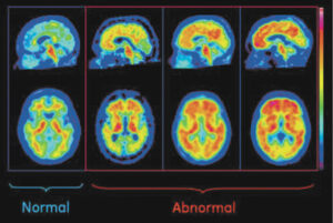
RAVE Imaging is proud to offer Vizamyl PET/CT brain scans, a powerful, FDA-approved tool for evaluating beta-amyloid plaque in adults experiencing cognitive impairment. Vizamyl imaging is specifically designed to assist in the evaluation of patients who are being assessed for Alzheimer’s disease or other causes of cognitive decline.
What sets Vizamyl apart is its ability to identify beta-amyloid plaques as soon as cognitive symptoms appear, enabling earlier intervention than ever before. Vizamyl is also the only FDA-approved amyloid imaging agent designed for color image interpretation. This distinction is significant: color imaging provides clearer, more precise differentiation between scans that are negative for amyloid plaque and those that show a positive amyloid plaque load. The result is greater diagnostic confidence for physicians and more actionable information for patients.
The Centiloid Advantage
RAVE Imaging holds a distinct advantage: it is the only facility in the area with the specialized software required to generate a Centiloid Value from Vizamyl scans. The Centiloid is a standardized, tracer-independent unit of measurement used to quantify the precise amount of beta-amyloid plaque present in the brain. This level of quantification goes beyond a simple positive or negative result, it gives clinicians a measurable data point to track disease progression, evaluate treatment response, and make more informed care decisions over time.
Expert Radiologists You Can Trust
RAVE’s Vizamyl PET/CT scans are interpreted by board-certified Neuroradiologists Dr. Neerav Mehta and Dr. Bradley Close, specialists with the expertise to deliver accurate, meaningful results. Their involvement ensures that every scan is read with the highest level of clinical precision.
If you or a loved one is experiencing memory loss or cognitive changes, don’t wait. Contact RAVE Imaging today to learn how a Vizamyl PET/CT scan could be the first step toward answers and a path forward.
941-488-7781
www.raverad.com
VENICE
512 S. Nokomis Ave
Venice, FL 34285
Hours: 8:00am-5:00pm
ENGLEWOOD
900 Pine Street
Englewood, FL 34223
Hours: 8:00am-5:00pm
SARASOTA
3501 Cattlemen Road
Sarasota, FL 34223
Hours: 8:00am-5:00pm