
By Dr. Stephen Pomeranz, Founder & CEO, ProScan Imaging
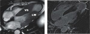
Hypertrophic Cardiomyopathy (genetically induced thick heart) with VS= ventricular septum and LW = Left “free” wall and an apical ventricular aneurysm, arrows.
Advanced imaging has assumed a major role in the diagnosis of heart disease. For instance, cardiac CT has received a class 1 (level of recommendation is strong) and evidence level A (randomized clinical trials yield the highest level of clinical evidence) for use in both stable and acute chest pain settings according to the American Heart Association and American College of Radiology chest pain guidelines.
Thus a targeted precision planned cardiac CT can greatly influence patient care and outcomes. The DISCHARGE trial (Diagnostic Imaging Strategies for Patients with Stable Chest Pain and Intermediate Risk of Coronary Artery Disease) randomized 3561 patients to either cardiac CT or invasive coronary angiography (ICA) as an initial testing strategy. No significant difference in major events was found 3.5 years later. Here are some relevant and timely facts.
• Heart disease is the leading cause of death for men and women in the U.S
• Cardiovascular diseases are the leading cause of death globally, taking an estimated 17.9 million lives each year
• Heart disease causes 2x as many deaths as cancer and causes more deaths than all the other causes of death combined.
The best way to approach heart disease is “not to get it” or, said another way, do what you can as early in life as you can, to prevent it.
In other words, the noninvasive CT tests performed similarly to the invasive angiography test.
Life’s Essential Preventative 8 Prevention tenets include Diet; Exercise; Sleep; Lipid Level; Weight; Blood Sugar; Blood Pressure; Smoking Avoidance
As an aside, the Mediterranean diet with generous amounts of virgin olive oil is the only diet shown to diminish heart disease.
ProScan at NCH now provides multiple state of the art “best in class” fast CAT (CT) scanners; cardiac PET and multiple cardiac capability MRI scanners.
Who should consider cardiac evaluation and/or screening in general:
• Smokers
• Family History of Heart Disease
• “Peace of Mind”
• Typical (HTN, Diabetes, Age, Hormones, Cholesterol, LDL-C, etc.) or Atypical labs like (Lp(a) or Factor V Leiden)
• Equivocal Other or Positive Stress Test
CT Cardiac Calcium Score
Atherosclerosis is the disease that affects the arteries that nourish the heart. When the “plaques” or ulcers work their way into the wall of these vessels, inflammation ensues. At this point, the inflammation may subside and “heal” producing calcium in the healing process. So, in a sense, these are the “good” plaques.
However, these calcified plaques serve as a biomarker or predictor of plaques that are inflamed, ulcerated and noncalcified. In other words, they haven’t healed. These soft plaques are made of fat and scar/ fibrous tissue. It is these lesions that may produce potential heart attack, chest pain, and other symptoms. They are known as “vulnerable” plaques.
The test must be interpreted based on patient age, gender, and history. A score of 5 in a 45-year-old woman is very significant. The same score in an 80-year-old is not.
FACT: Lowering LDL-c by 38.7 mg/dL and lowering SBP by 10 mmHg results in a relative risk reduction of major CV events of 78%
The results can be divided into nominal, mild, moderate and severe levels:
Nominal: 1-10
Mild: < 100
Moderate: 100-400
Severe: Greater than 400
For any positive score, consult your physician, concierge doctor or cardiologist. If the test is positive, you have several diagnostic options discussed below. Many physicians will look at the cardiac score as a determinant of whether their patient should be aggressively treated with statins and other cholesterol lowering medications.
Cardiac CT Angiography (CCTA). Normal Study
The CCTA is a test that uses a CAT scan (CT) and an injection of iodinated contrast to make an angiogram like image of the heart that shows the coronary arteries heart and aorta. It “sees” the nonhealed/noncalcified plaques. The plaques are visually graded based on depth, percent narrowing, shape and even length. If the narrowing is significant based on the interpretation there are several options:
– Applying AI to your CCTA (with CLEERLY; or HEARTFLOW) Cardiology consultation
– Cardiac PET scanning
CLEERLY (artificial intelligence)
Cleerly is an artificial intelligence (AI) technique which analyzes the CCTA retrospectively. It looks with a computer at plaque: Composition; volume of plaque; shape; length; width; depth. In other words; it creates a “google map” of the coronary arteries that nourish the heart.
With regard to “type of plaque”, the SCOT-Heart (Scottish CT of the Heart) trial showed that low-attenuation plaques resembling fat or vulnerable plaque was a stronger predictor of heart attack than: stenosis severity; coronary calcium; or cardiac risk score calculation on a risk calculator model.
HEARTFLOW (artificial intelligence)
Once the CCTA has been performed, the areas of narrowing are assessed by the reader in each major coronary vessel. However, with some areas of narrowing, it may be difficult to assess whether the narrowing is significant or, in other words, compromises the flow of blood to the heart muscle downstream. Heartflow is an AI algorithm that looks at how fast the contrast appears downstream to an area of narrowing. In calculating this, the AI can tell if the narrowing of rate is flow limiting, thereby raising awareness and aggressiveness in diagnosis and management. Simply stated, if the number applied to the vessel downstream is < 0.8, than the narrowing is significant and requires additional attention. This patients heart flow was negative downstream from the narrowing.
Cardiac PET (positron emission tomography)
Cardiac PET is phasing in as a higher quality, faster, safer tool that is replacing the cardiac stress test. It uses a radioactive injection of a substance called Rubidium that has to be delivered in a special generator each day.
Cardiac PET is:
– 9X faster than regular stress testing
– 7X lower radiation dose
– More accurate
– Has fewer artifacts
The study looks at both the overall circulation to the heart as well as for microvascular dysfunction. This can help identify disease early on, allowing physicians to employ medical therapy for improved prevention of myocardial ischemic events. Stress and rest imaging of the heart takes place in only 20 minutes, much shorter than the standard SPECT scan that takes 3-4 hours. This test can be used to decide if the patient needs medical therapy or interventional cardiology consultation.
Simply stated, Cardiac PET is a superior diagnostic test to standard stress testing.
Cardiac Magnetic Resonance Imaging (MRI)
Hypertrophic Cardiomyopathy (genetically induced thick heart) with VS= ventricular septum and LW = Left “free” wall and an apical ventricular aneurysm, arrows.
ProScan at NCH now provides cardiac MRI at three locations in Collier County (Ninth Street, Sierra Meadows and Crosspointe). MRI or magnetic resonance imaging provides anatomic, functional, and blood supply information about the heart. The radiology/ cardiology interpreting team has deep experience for almost 2 decades in performing and reading cardiac MR (as well as CT). The applications of MRI of the heart have been regularly expanding and is now used to diagnose a wide variety of cardiovascular conditions.
MR delivers:
– No radiation
– Usually requires a small volume of injection
– Takes approximately 45 mins – 1 hour to perform
Some indications for cardiac MRI include:
– Congenital heart disease
– Inflammation or myocarditis (as in COVID)
– Evaluation for causes of an enlarged or “thick” heart
– Evaluate the cause of decreased heart function
– To determine the cause of an arrhythmia
– To determine whether and how much muscle damage has occurred from prior heart attack or other various disease
– Tumors and masses
For more information or to schedule an appointment, call 239-624-4443 www.proscan.com
Stephen J. Pomeranz, M.D.
Founder & CEO, ProScan Imaging
Dr. Pomeranz is renowned as a world leader in the field of diagnostic imaging, particularly MRI. His experience interpreting hundreds of thousands of MRI examinations (including MSK, Neuro, Body, Breast and Prostate studies) and his passion for education and mentoring come together to provide an excellent learning experience for physicians and healthcare professionals.
Dr. Pomeranz has written, co-authored, or edited numerous MRI texts, including the essential three-volume reference The MRI Total Body Atlas, as well as many multimedia teaching materials. He is also the founder of MRI Online, the world’s largest collection of online MRI Education videos available for Continuing Medical Education (CME) credits.