By Myles Rubin Samotin, MD – Board Certified Orthopaedic Surgeon, Fellowship Trained in Foot and Ankle –


As most of us know, a tendon is an extension of a muscle and attaches itself to a bone, allowing us to use our muscles to move our bones and joints over and over again. The posterior tibial muscle becomes a tendon on the inside (medial side) of our lower leg. It then runs behind the inside ankle (medial malleous) and attaches to eight bones in the midfoot. Since all the attachments sit in the middle of the arch, this muscle and tendon help support the foot arch.
Tendons are made up of collagen fibers sort of like a rope. As we get older, tendons become worn, with individual fibers becoming inflamed, calcifying or even rupturing. This leads to the tendon weakening. As the tendon tries to heal itself from this wear and tear, scar tissue may form into a knot or nodule within the tendon called tendonosis. This area is weaker than the tendon itself and can eventually lead to a tendon rupture. If the larger area of tendonosis becomes inflamed, it becomes tendonitis.
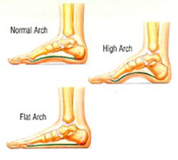
Like most aging muscles and tendons, the posterior tibial tendon will start to wear and lose elasticity, causing the arch of the foot to start to flatten. BUT, we use this tendon with EVERY STEP WE TAKE!!! We cannot stop using it, since it is very important in ambulation. So the tendon will continue to wear and will continue to lose its ability to support the arch and the foot.
As the tendon continues to degenerate, stretch out and lose its flexibility, the foot will become flatter and flatter. Even in a short time the actual shape of the foot will change significantly. This can result in changes in ligaments, bones and tendons of the foot and the results can lead to a very painful foot.
The symptoms of tendonosis/tendonitis of the posterior tibial tendon may include pain and swelling on the inside of the ankle or midfoot, loss of the foot arch and the development of flatfoot. Other symptoms might include weakness and an inability to stand on ones toes, and/or tenderness over the midfoot, especially when under stress during physical activity. However, not everyone may have symptoms and flatfoot may continue to progress without the patient being aware of the changes occurring in his or her foot. It is for this reason that a new onset or one that is getting worse should always be evaluated by an orthopaedic foot and ankle specialist to determine if any changes have occurred in your foot.
Of course, not everyone develops a posterior tibial tendon problem. However, there are people who are more at risk than others. This tendinopathy often occurs more commonly in women, especially those over the age of 50, and may be due to an inherent abnormality of the tendon. Also people who are obese, who have diabetes, who suffer from hypertension or who have had previous surgery or trauma to the inner side of the foot may be more prone to suffering from this tendinopathy. Some inflammatory diseases, such as rheumatoid arthritis can harmfully affect the tendon as well.
What happens if I start to suffer from these symptoms? Proper diagnosis should be made by a foot and ankle specialist, especially one who understands all the concepts of bones, such as an orthopaedic foot and ankle specialist. Having the expertise to understand the problem will allow the M.D. to per- form a special physical examination which will allow him to make the proper diagnosis. Imaging tests such as X-Ray and MRI will be performed to assess the extent of damage.
Treatment of posterior tibial tendinopathy may be done conservatively. Possibilities include the usage of non-steroidal anti-inflammatories drugs, casting or placing the affected limb into a boot. The doctor may also place heel wedges or arch supports into your shoes. He may also prescribe custom orthotics for you to wear to treat early minimal symptoms. Physical therapy may be used to try and strengthen the tendon. However, as I stated earlier, symptoms do not always show up until the deformity from the tendinopathy is much more advanced and resulting treatments are much more involved.
Conservative treatments may also not provide any relief from your symptoms. Fortunately, as an orthopaedic fellowship trained foot and ankle specialist, my expertise gives me several surgical alternatives, depending on whether I am dealing with a tendonosis, a tendonitis, a partially or fully ruptured tendon, or changed foot anatomy caused by a bad posterior tibial tendon. This may include a simple procedure such as a Synovectomy, where the tendon is debrided (cleaned) and any bad or inflamed tissue will be removed. Other possibilities also are a tendon transfer which uses another tendon to help the posterior tibial tendon. I may also do a bone realignment, an Osteotomy, which will help with feet that have changed their anatomy or architecture from severe posterior tibial tendinopathy.
If the changes in the foot are allowed to progress due to posterior tibial tendinopathy, eventually the feet will get flatter and flatter and the result will be that the bones in the middle of the foot will change how they work with other bones. This could lead to severe arthritic changes in these joints and walking and even standing could become extremely painful. If the arthritis becomes bad enough, the only way to rid the symptoms of the pain would be through a fusion of these bones, known as an Arthrodesis. This is a very effective operation to eliminate or lessen the pain problem as well as provide stability for walking.
What is the best thing you can do for yourself? If you have any symptoms, the best way, as I said, is to have your feet evaluated by the proper Orthopaedic specialist. This is a problem that often worsens over time with treatment becoming more and more complicated. With 28 bones in your foot, you need to be evaluated by a Board Certified Orthopaedic Surgeon with a Sub-specialty, Fellowship Trained in Foot & Ankle surgery. In fact I am the only surgeon with these qualifications in our area. I believe this makes me uniquely able to deal with these problems in a state-of-the-art atmosphere and method that will keep you in good hands and provide you with the most desired result.
Myles Rubin Samotin, MD
Board Certified Orthopaedic Surgeon Fellowship Trained, Sub-specialist in Foot and Ankle Surgery Columbia University, Maimonides Medical Center, Hospital for Joint Diseases, New York City
941.661.6757
713 E. Marion Ave, Suite 135 (3rd Floor),
Punta Gorda, FL 33950
 Southwest Florida's Health and Wellness Magazine Health and Wellness Articles
