By Haris Turalic, MD, F.A.C.C. –
 Heart disease is the leading cause of death and a major cause of disability in the U.S. At Lehigh Regional Medical Center, we provide comprehensive, technologically advanced heart care delivered by knowledgeable, experienced cardiac professionals. Our heart specialists offer state-of-the-art heart care, from diagnosis and emergency intervention to the latest treatments, preventive education and rehabilitation.
A cardiologist’s most important resource is the stress test when a diagnosis needs to be made to determine the existence of heart disease. There are several different tests; and each test has it’s own individual and highly specific requirements. Not only do the specific tests involved influence the reliability of the diagnosis but also the conditions of the testing as well. One thing doctors want to discover is just how much stress the heart can take before it impacts the behavior or functionality of the heart.
The following discusses the benefits of the two main types of cardiovascular stress testing: standard exercise stress test and imaging stress test.
Exercise Stress Test
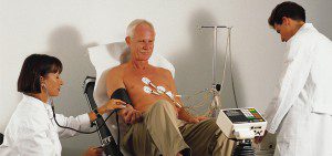
The exercise stress test, while best suited for a specific application, is the one that most people are familiar with. A standard exercise stress test uses an electrocardiogram (EKG) to detect and record the heart’s electrical activity.
An EKG shows how fast your heart is beating and the heart’s rhythm (steady or irregular). It also records the strength and timing of electrical signals as they pass through your heart.
During a standard stress test, your blood pressure will be checked. You also may be asked to breathe into a special tube during the test. This allows your doctor to see how well you’re breathing and measure the gases that you breathe out.
A standard stress test shows changes in your heart’s electrical activity. It also can show whether your heart is getting enough blood during exercise.
Imaging Stress Test
As part of some stress tests, pictures are taken of your heart while you exercise and while you’re at rest. These imaging stress tests can show how well blood is flowing in your heart and how well your heart pumps blood when it beats.
One type of imaging stress test involves echocardiography (echo). This test uses sound waves to create a moving picture of your heart. An exercise stress echo can show how well your heart’s chambers and valves are working when your heart is under stress.
A stress echo also can show areas of poor blood flow to your heart, dead heart muscle tissue, and areas of the heart muscle wall that aren’t contracting well. These areas may have been damaged during a heart attack, or they may not be getting enough blood.
Other imaging stress tests use radioactive dye to create pictures of blood flow to your heart. The dye is injected into your bloodstream before the pictures are taken. The pictures show how much of the dye has reached various parts of your heart during exercise and while you’re at rest.
Tests that use radioactive dye include a thallium or sestamibi stress test and a positron emission tomography (PET) stress test. The amount of radiation in the dye is considered safe for you and those around you. However, if you’re pregnant, you shouldn’t have this test because of risks it might pose to your unborn child.
Imaging stress tests tend to detect heart disease better than standard (non-imaging) stress tests. Imaging stress tests also can predict the risk of a future heart attack or premature death.
An imaging stress test might be done first (as opposed to a standard exercise stress test) if you:
• Can’t exercise for enough time to get your heart working at its hardest. (Medical problems, such as arthritis or leg arteries clogged by plaque, might prevent you from exercising long enough.)
• Have abnormal heartbeats or other problems that prevent a standard exercise stress test from giving correct results.
• Had a heart procedure in the past, such as coronary artery bypass grafting or angioplasty and stent placement.
What Does Stress Test Show?
A standard exercise stress test uses an EKG to monitor changes in your heart’s electrical activity. Imaging stress tests take pictures of blood flow throughout your heart. They also show your heart valves and the movement of your heart muscle.
Doctors use both types of stress tests to look for signs that your heart isn’t getting enough blood flow during exercise. Abnormal test results may be due to coronary heart disease or other factors, such as poor physical fitness.
If you have a standard exercise stress test and the results are normal, you may not need further testing or treatment. But if your test results are abnormal, or if you’re physically unable to exercise, your doctor may want you to have an imaging stress test or other tests.
Even if your standard exercise stress test results are normal, your doctor may want you to have an imaging stress test if you continue having symptoms (such as shortness of breath or chest pain).
Imaging stress tests are more accurate than standard exercise stress tests, but they’re much more expensive.
Imaging stress tests show how well blood is flowing in the heart muscle and reveal parts of the heart that aren’t contracting strongly. They also can show the parts of the heart that aren’t getting enough blood, as well as dead tissue in the heart, where no blood flows.
If your imaging stress test suggests significant coronary heart disease, your doctor may want you to have more testing and treatment.
Stress tests are generally performed to evaluate if chest pains are related to heart problems, to determine is arteries have blockages, to identify any irregular heart rhythm, to monitor the heart’s response to treatments and procedures, to determine a safe level of physical activity, and to plan rehabilitation after a heart attack.
If you don’t have chest pain when you exercise but still get short of breath, your doctor may recommend a stress test. The test can help show whether a heart problem, rather than a lung problem or being out of shape, is causing your breathing problems.
Performing periodic stress tests can be a useful way of monitoring the functionality of the heart and can track progress of patients with existing heart disease. For more information about the benefits of the various types of stress tests available, contact the Lehigh Regional Cardiovascular team by calling 239-368-6017. Don’t wait until it is too late!


