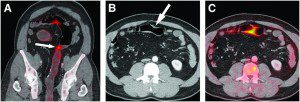
 Until recently, diagnosis of small bowel pathology had relied primarily on radiological techniques, in part due to the relative inaccessibility of the small bowel to conventional endoscopy. Progress continues to be made in cross-sectional imaging technologies, harnessing the power of computer tomography, MRI and ultrasound, facilitating rapid, accurate and minimally invasive investigation of the small bowel and adjacent tissues.
What is CT Enterography?
Computed tomography, more commonly known as a CT or CAT scan, is a diagnostic medical test that, like traditional x-rays, produces multiple images or pictures of the inside of the body.
CT enterography is a special type of computed tomography (CT) imaging performed with intravenous contrast material after the ingestion of liquid contrast material that helps produce high resolution images of the small intestine in addition to the other structures in the abdomen and pelvis.
CT enterography is a non-invasive imaging technique that offers superior small bowel visualisation compared with standard abdomino-pelvic CT, and provides complementary diagnostic information to capsule endoscopy and MRI enterography. CT enterography is well tolerated by patients and enables accurate, efficient assessment of pathology arising from the small bowel wall or surrounding organs.
The cross-sectional images generated during a CT scan can be reformatted in multiple planes, and can even generate three-dimensional images. These images can be viewed on a computer monitor, printed on film, or transferred to a CD or DVD.
CT images of internal organs, bones, soft tissue and blood vessels typically provide greater detail than traditional x-rays, particularly of soft tissues and blood vessels.
Physicians use CT Enterography to identify and locate:
• small bowel inflammation
• bleeding sources within the small bowel
• small bowel tumors
• abscesses and fistulas
• bowel obstruction
CT enterography is also used to diagnose Crohn’s disease, and determine its location, severity and unexpected complications, in order to guide effective treatment.
Benefits of CT Enterography
• CT scanning is painless, noninvasive and accurate.
• A major advantage of CT is its ability to image bone, soft tissue and blood vessels all at the same time.
• Unlike conventional x-rays, CT scanning provides very detailed images of many types of tissue as well as the lungs, bones, and blood vessels.
• CT examinations are fast and simple; in emergency cases, they can reveal internal injuries and bleeding quickly enough to help save lives.
• CT has been shown to be a cost-effective imaging tool for a wide range of clinical problems.
• Compared to other imaging procedures of the small intestine, CT enterography is able to visualize the entire thickness of the bowel wall and to evaluate surrounding soft tissues. The other examinations, some of which are invasive, are only able to image the inner lining of the small intestine.
• CT enterography has been shown to diagnose and/or rule out certain conditions/diseases that could help determine your future medical care.
• CT enterography may eliminate the need for video capsule endoscopy (VCE) and the potential complications of that procedure.
• CT enterography allows other organs in the abdomen to be seen.
• CT is less sensitive to patient movement than MRI.
• CT can be performed if you have an implanted medical device of any kind, unlike MRI.
• No radiation remains in a patient’s body after a CT examination.
Who interprets the results?
A radiologist with expertise in supervising and interpreting radiology examinations will analyze the images and send an official report to your primary care physician or physician who referred you for the exam, who will discuss the results with you.


