 As technology advances, understanding medical exams and procedures becomes more complex. The quality of services provided is an important consideration.
The American Cancer Society endorses mammography, along with yearly physical examinations and monthly self-examinations, as the most effective means of detecting breast cancer at its earliest and most treatable stage. Generally, mammography can reveal benign and cancerous growths before you or your physician can feel them. If detected at the earliest stage, breast cancer has a five-year survival rate of over 95 percent, as small breast cancers are more treatable and can be removed before they spread to other parts of the body.
Breast cancer is the most common form of cancer in American women. Unfortunately, 70% of women have no identifying risk factors. The American Cancer Society recommends mammography as a life saving tool for screening women without symptoms for breast cancer. And 3D Mammography specifically is becoming the preferred choice for physicians in Southwest Florida. With over 30 years of experience and 10 Board Certified Radiologists, Radiology Associates of Venice & Englewood (RAVE) is proud to offer 3D Mammography to our patients.
What is 3D Mammography?
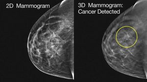
3D mammography is a revolutionary state of the art technology approved by the FDA in February 2011, which gives radiologists the ability to view inside the breast layer by layer, helping to see the fine details more clearly by minimizing overlapping tissue. During a 3D mammogram, multiple low-dose images known as “slices” of the breast are acquired at different angles. With 3D technology, the radiologist can view a mammogram in a way never before possible.
Is 3D a separate exam or part of my usual mammogram?
The 3D exam is a separate procedure that is performed at the same time as your regular mammogram.
What is the cost and will my insurance cover the 3D exam?
Medicare does cover 3D mammography. Even though 3D mammography is FDA approved and covered by Medicare, most private insurance companies are not yet reimbursing for this exam. However, RAVE has never charged the patient the additional 3D portion of the exam if their insurance doesn’t cover it.
“The Radiologists of RAVE include the additional 3D imaging regardless of payment because it’s in the best interest of patient care, so there is never an additional charge.”(Philip Mihm, M.D. RAVE Radiologist)
What are the benefits?
FEWER MAMMOGRAM CALLBACKS for additional mammography – 3D mammography helps distinguish harmless abnormalities from real cancers, leading to fewer callbacks for additional mammography and less anxiety for women. With 3D mammography, RAVE radiologists have reduced patient callback rates by 20-30 percent.
Doctors and scientists agree that early detection is the best defense against breast cancer. 3D mammography has been shown in clinical studies to be more accurate than conventional mammography alone by detecting cancers earlier. This new technology increases breast cancer detection by 38%. It’s truly an important component in the screening process.
After 3D Mammography, if continued tests and imaging are needed, RAVE uses state-of-the-art technology, including MRI guided breast biopsies and the Philips 3T wide bore MRI that allows our radiologists to view the breasts in a higher resolution, enabling us to have even more clarity within the breasts. RAVE has been performing MRI breast imaging for over 15 years and with Wide Bore technology, it allows us to accommodate most any sized patient comfortably. With the Philips 3T wide bore MRI, we are able to cut down on the amount of time it takes for the patient to be scanned. Most Breast MRI’s take 30 minutes or less, allowing the patient to go on with their day with little disruption.
How long will it take?
The exam will take about 4 seconds longer per view while in compression than the 2D mammography.
How much radiation will I be exposed to?
It varies from person to person and is roughly equivalent to film/screen mammography. The amount of radiation is below government safety standards.
What if my doctor did not mention 3D Mammography to me?
3D is an optional service at this time and elected by the patient. Many physicians know about our new 3D technology and the feedback we have received has been very positive. If you need additional information to help you make this decision, please visit www.RaveRad.com.
Why is RAVE Radiology offering 3D Mammography?
RAVE prides itself on offering the highest quality care for our patients. Our radiologists believe strongly that 3D mammography will benefit our patients.
We are approaching our 3rd Breast Cancer Awareness month since the COVID pandemic began. Breast Imaging, usually fairly insulated from worldly events, has shared in the challenges over the past few years. Initially concerns regarding post vaccination lymphadenopathy made its way to the nightly news. Confusion set in about whether and when to get a mammogram following vaccination. Luckily this was never a diagnostic dilemma for us at RAVE and we were able to encourage most women to stick to their annual screening schedule. Unfortunately, and for understandable reasons, several women have not come in for mammographic screening since the pandemic begin. Because breast cancer detection and management are a primary mission at RAVE, we have risen to the challenge of ensuring safe access to breast cancer screening exams and any additional/follow-up care needed. Please be reassured that we are providing our standard high level of imaging care while maintaining/exceeding current CDC guidelines to ensure patient safety.
Furthermore, it’s worth noting that RAVE offers the cutting edge in imaging technology unsurpassed in our region. We utilize the newest mammographic machines, each equipped with 3D Intelligent HD Clarity from Hologic. Tradename aside, the image quality is unparalleled, akin to the highest end Ultra HD television. This is important not only because it allows us to diagnose smaller cancers but also facilitate accurate characterization of benign findings other radiology groups mistake for malignancy.
Our ultrasound equipment is also the highest quality available in the industry which has implications for our breast cancer mission as well as our other imaging services. Finally, our 3 Tesla MRI also generates extremely high-quality breast images which facilitate screening in our high-risk patients and important staging information in our women diagnosed with breast cancer. Equipped with these tools we recently identified a 3mm cancer via mammography! I would argue this tiny cancer is the earliest and smallest lesion a screening examination could hope to accurately identify.
We do not stop at the detection of breast cancer! Currently we are providing ultrasound breast biopsies at our Venice and Sarasota offices. At RAVE we know biopsy procedures are a scary process. We work hard to inform our patients beforehand regarding what to expect during the procedure. Professional, personalized, warm, and caring treatment is provided during the procedure. Lastly, follow up afterwards ensures nothing falls through the cracks. Most women leave our biopsy suite much more informed and prepared regarding their individual case and the forthcoming steps. For our referring physicians we provide critical radiology pathology concordance following all biopsies to help manage pathological results they may not be familiar with. This ensures suspicious lesions are pursued even if pathology results are not as expected and offers reassurance when benign results match less suspicious findings. In the not-too-distant future we will be offering the newest biopsy method which allows sampling of “3D” or tomosynthetic findings. This system is the final piece in the definitive management of the lesions we can detect and complements our current ability to perform MRI guided breast biopsies. I am very proud to be a part of the comprehensive breast program we offer at RAVE and am very grateful for the opportunity to serve our area’s patients and referring physicians.
RAVE is excited to announce that we will be providing a more advanced DEXA Bone Density study at all three locations. DEXA with TBS.
What is the difference between DEXA scan and DEXA scan and TBS?
Bone mineral density measured by DEXA provides information regarding the quantity of the mineral bone only. TBS is a measurement of bone quality. Using both together gives the practitioner a better picture of the bone strength of an individual patient.
Ask your health care provider for DEXA with TBS for a better understanding of your bone fracture risk.
Radiology Associates of Venice & Englewood
VENICE
512-516 S. Nokomis Ave
Venice, FL 34285
941-488-7781
Hours: 8:00am-5:00pm
ENGLEWOOD
900 Pine Street
Englewood, FL 34223
941-475-5471
Hours: 8:00am-5:00pm
SARASOTA
3501 Cattlemen Road
Sarasota, FL 34223
941-342-RAVE (7283)
Hours: 8:00am-5:00pm


