– By Advanced Imaging of Port Charlotte –


When PKD causes kidneys to fail, which usually happens after many years, the patient requires dialysis or kidney transplantation. PKD can also cause cysts in the liver and problems in other organs, such as blood vessels in the brain and heart. In the United States, about 600,000 people have PKD, and cystic disease is the fourth leading cause of kidney failure. Autosomal dominant PKD is the most common inherited disorder of the kidneys. The phrase “autosomal dominant” means that if one parent has the disease, there is a 50 percent chance that the disease gene will pass to a child. In some cases, this disease occurs spontaneously. In these cases, there is not a genetic component in the development of PKD. With autosomal PKD symptoms may not appear until the patient is older. When the symptoms begin, they are due to cysts that grow in the nephrons, which are the filtering devices inside the kidneys. As the cysts grow, they cause the kidneys to become enlarged. This can cause other symptoms such as high blood pressure and pain. Initially the pain will appear in the back and sides. In addition, the patient may experience headaches that can be attributed to the high blood pressure and potential aneurysms in the brain.
PKD is usually diagnosed by kidney imaging studies. The most common form of diagnostic kidney imaging is ultrasound, however, computerized tomography (CT) scans or magnetic resonance imaging (MRI) are increasingly used to diagnose the disease due to their ability to detect cysts and other physiological changes.
Once cysts have grown to about one-half inch, however, diagnosis is possible with imaging technology. Ultrasound, which passes sound waves through the body to create a picture of the kidneys, is used most often. Ultrasound imaging does not use any injected dyes or radiation and is safe for all patients, including pregnant women. It can also detect cysts in the kidneys of a fetus, but large cyst growth this early in life is uncommon in autosomal dominant PKD. An ultrasound can measure the size and appearance of the kidneys and detect tumors, congenital anomalies, swelling and blockage of urine flow.
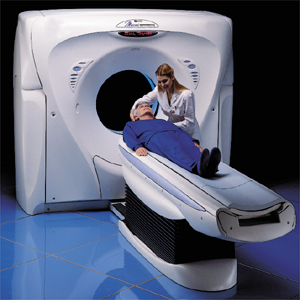
Computerized Tomography (CT) uses a computer to reconstruct multiple x-ray data samples. It is best used to detect kidney stones or tumors. It can assess most of the details similar to ultrasound, but CT’s are sometimes done with contract, intravenous (IV) contrast dye is used. Some people may have an allergic reaction to the contrast agent so it is important that the patient give an accurate history of any allergies, especially to shellfish or contrast agents.
A magnetic resonance angiogram (MRA) is a type of magnetic resonance imaging (MRI) scan that uses a magnetic field and pulses of radio wave energy to provide pictures of blood vessels inside the body. In many cases MRA can provide information that can’t be obtained from an X-ray, ultrasound, or computed tomography (CT) scan. MRA can find problems with the blood vessels that may be causing reduced blood flow. With MRA, both the blood flow and the condition of the blood vessel walls can be seen. The test is often used to look at the blood vessels that go to the brain, kidneys, and legs. A magnetic resonance angiogram (MRA) is done to look for narrowing (stenosis) of the blood vessels leading to the lungs, kidneys, or legs.
Advanced Imaging offers the highest quality and latest technology imaging available to diagnose kidney disease at its earliest stage. Although a cure for autosomal dominant PKD is not available, treatment can ease symptoms and prolong life. If you have a history of family kidney disease or exhibit any of the symptoms of kidney disease, contact your doctor who will be able to determine what imaging and treatment are best for you.
Advanced Imaging of Port Charlotte
941-235-4646
www.advimaging.com
 Southwest Florida's Health and Wellness Magazine Health and Wellness Articles

