 Southwest Florida's Health and Wellness Magazine Health and Wellness Articles

 Two men every five minutes are diagnosed with prostate cancer, the second leading cause of cancer death in men. According to the American Cancer Society, an estimated 220,000 new cases of prostate cancer are diagnosed each year, and nearly 35,000 men die annually from the disease.
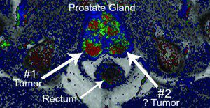
MRI is a reliable tool for possible early detection of prostate cancer and other prostate-related conditions. MRI uses radiofrequency waves to create a detailed cross-sectional image of the prostate and surrounding tissues.
Prostate MRI uses advanced magnetic resonance imaging to create very accurate and clear images of the prostate gland. These images are diagnostic quality and can be useful when diagnosing possible prostate diseases. Medical images resulting from prostate MR can be combined with powerful post-processing computer programs to provide detailed information about the prostate. This information can offer a wider variety of diagnosis and treatment options for clinicans and patients. If you feel you could benefit from a prostate MRI please discuss with your physician.
Images provided from MR, however, do not always indicate cancer. Prostate MR images identify specific regions of the gland that may appear suspicious and can be further evaluated through a targeted MR-guided biopsy procedure (also available at Advanced Imaging of Port Charlotte). MRI of the prostate can also be used to evaluate other prostate conditions, including prostatitis (inflam-mation of the prostate) and benign prostatic hyperplasia (BPH)(enlargement of the prostate)
MR-guided biopsies may be helpful to patients who have had several sessions of TRUS-guided biopsies with negative results. Compared to published cancer yield rates of up to 15% greater with their TRUS-guided biopsies, the increased rate of detection may give greater confidence to both physicians and patients.
What to Expect from the MR Imaging Process
An MRI is a non-invasive and painless medical procedure used to produce accurate, detailed pictures of organs and tissues to diagnose a variety of medical conditions. When receiving an MRI, patients are positioned on a moveable examination table, and in some cases, straps and bolsters are used to help patients remain still and in the correct position during imaging. Small devices that contain coils capable of sending and receiving radio waves, may then be placed around or adjacent to the area of the body that is being studied.
The patient is then moved into the magnet of the MRI unit. The radiologist and the technologist will leave the room while the MRI examination is performed on the patient. When the examination is completed, patients may be asked to wait until the technologist or radiologist checks the images, in the event additional images are required.
MRI exams generally include multiple runs (sequences), some of which may last several minutes. The entire exam is usually completed in 15-45 minutes.
For more information about the prostate MRI and other diagnostic imaging services available at Advanced Imaging call 941-235-4646 today.
Benefits:
• Prostate MRI is a noninvasive imaging technique that does not require exposure to ionizing radiation.
• Prostate MRI provides clear and detailed images of the soft-tissue structures of the prostate that may not be assessed adequately with other imaging methods such as x-ray, ultrasound or computed tomography (also called CT or CAT scanning). The detail makes MRI a helpful tool in early diagnosis and evaluation of tumors.
• Prostate MR images can help physicians evaluate the function as well as the structure of many organs.
• MRI contrast material is less likely to produce an allergic reaction than the iodine-based materials used for conventional x-rays and CT scanning.