 Southwest Florida's Health and Wellness Magazine Health and Wellness Articles

 Urography uses imaging and contrast material to evaluate or detect blood in urine, kidney or bladder stones, and cancer in the urinary tract. Urography with conventional x-ray is known as intravenous pyelogram (IVP). Urography may also be performed using computed tomography (CT). CT urography is painless and proven effective in detecting urinary tract issues.
What is Urography?
Urography is an examination used to evaluate the kidneys, ureters and bladder. Excretory urography, also known as intravenous pyelogram, is performed using conventional x-ray after the intravenous administration of radiographic contrast material. This technique is still performed for pediatric patients and for younger adult patients.
Computed tomography (CT) urography uses intravenous contrast material to obtain images of the urinary tract. CT urography (CTU) is used as primary imaging techniques to evaluate patients with blood in the urine (hematuria), follow patients with prior history of cancers of the urinary collecting system and to identify abnormalities in patients with recurrent urinary tract infections. In addition to imaging the urinary tract, CT urography can provide valuable information about other abdominal and pelvic structures and diseases that may affect them.
What are some common uses of the procedure?
Urography images are used to evaluate issues or detect abnormalities in portions of the urinary tract, including the kidneys, bladder and ureters, including:
• Hematuria (blood in urine)
• Kidney or bladder stones
• Cancers of the urinary tract
Exam Preparation
• No Barium.
• No solid food 4 hours prior to the exam.
• Drink water, there is no restriction on your waterintake.
• If you have an allergy to the IV iodinated contrast media you will need to be pre-medicated. Click here to view the Iodine Contrast Media Preparation Guide.
• Patients may be asked to dress into an exam gown.
During the Exam
• Any metal that is within the area to be scanned will need to be removed.
• The technologist will insert an IV for the injection of the iodinated contrast media.
• During the scan your arms will be brought above your head and a compression belt will be placed around your waist. Pressure will be applied for a short time during the scan.
• You will be asked to hold your breath for 10-20 seconds while the images are being taken.
• Exam should be completed within 15 minutes, although it could take longer.
After the Exam
• Following your exam you will be asked to remain in the department until your IV has been removed and a standard x-ray may be needed 20 minutes after the CT scan.
• Report will be forwarded to your doctor
Women should always inform their physician and the CT technologist if there is any possibility that they are pregnant. See the Safety page for more information about pregnancy and x-rays.
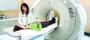
How does the procedure work?
CT scanning combines special x-ray equipment with sophisticated computers to produce multiple images or pictures of the inside of the body. These cross-sectional images of the area being studied can then be examined on a computer monitor, printed or transferred to a CD.
Modern CT scanners are so fast that they can scan through large sections of the body in just a few seconds, and even faster in small children. Such speed is beneficial for all patients but especially children, the elderly and critically ill, all of whom may have difficulty in remaining still, even for the brief time necessary to obtain images.
For children, the CT scanner technique will be adjusted to their size and the area of interest to reduce the radiation dose.
Benefits of CT Urography
CT urography has been proven effective in detecting issues or abnormalities in parts of the urinary tract including the kidneys, bladder and ureters, or as a follow-up test to further examine for recurrent or new cancers of the urinary tract.
• Compared to other imaging tests, CT urography provides superior anatomic detail of the urinary tract and surrounding structures.
• CT scanning is painless, noninvasive and accurate.
• A major advantage of CT is its ability to image bone, soft tissue and blood vessels all at the same time.
• Unlike conventional x-rays, CT scanning provides very detailed images of many types of tissue as well as the lungs, bones, and blood vessels.
• CT examinations are fast and simple; in emergency cases, they can reveal internal injuries and bleeding quickly enough to help save lives.
• CT has been shown to be a cost-effective imaging tool for a wide range of clinical problems.
• CT is less sensitive to patient movement than MRI.
• CT can be performed if you have an implanted medical device of any kind, unlike MRI.
• CT imaging provides real-time imaging, making it a good tool for guiding minimally invasive procedures such as needle biopsies and needle aspirations of many areas of the body, particularly the lungs, abdomen, pelvis and bones.
• A diagnosis determined by CT scanning may eliminate the need for exploratory surgery and surgical biopsy.
• No radiation remains in a patient’s body after a CT examination.
239-593-4222
www.NaplesImaging.com